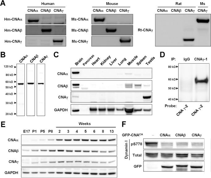
FIGURE 1.
CNAγ is expressed in the brain and dephosphorylates synaptic calcineurin substrates. A, human (Hm) and mouse (Ms) CNAα, CNAβ, and CNAγ and rat (Rt) CNAγ recombinant proteins were generated in 293T cells. Protein extracts were probed on a Western blot with anti-human CNAα, CNAβ, and CNAγ antisera; anti-mouse CNAα, CNAβ, and CNAγ antisera; and anti-rat CNAγ antiserum to demonstrate antibody specificity. For the rat CNAγ antiserum analysis, mouse CNAα and CNAβ recombinant proteins were used because of complete amino acid sequence conservation. The higher molecular weight of the mouse relative to the rat CNAγ protein is due to an N-terminal cycle 3 GFP tag present on the recombinant mouse form of the protein. B, hippocampus protein extract from 5-week-old mice was probed on a Western blot with anti-mouse CNAα, CNAβ, and CNAγ antisera. C, Western blotting was performed on mouse tissue protein extracts using anti-mouse CNAα, CNAβ, and CNAγ antisera and a GAPDH antibody. The lack of a Western blot signal in the pancreas tissue extracts is likely due to protein degradation. D, immunoprecipitation (IP) of CNAγ from a human neocortex sample. Purified antibodies against different regions of the protein were used to immunoprecipitate (γ-1) and probe for human CNAγ (γ-2). E, Western blot showing the expression of CNAα, CNAβ, and CNAγ (using custom antisera) and GAPDH in neocortex protein samples from mice at the indicated ages. E, embryonic day; P, postnatal day. F, 293T cells were co-transfected with GFP-tagged constitutively active versions of CNAα, β, or γ (GFP-CNACA) and a dynamin I expression construct. Western blotting was performed on protein extracts for Ser(P)-778 and total dynamin I and for GFP.

