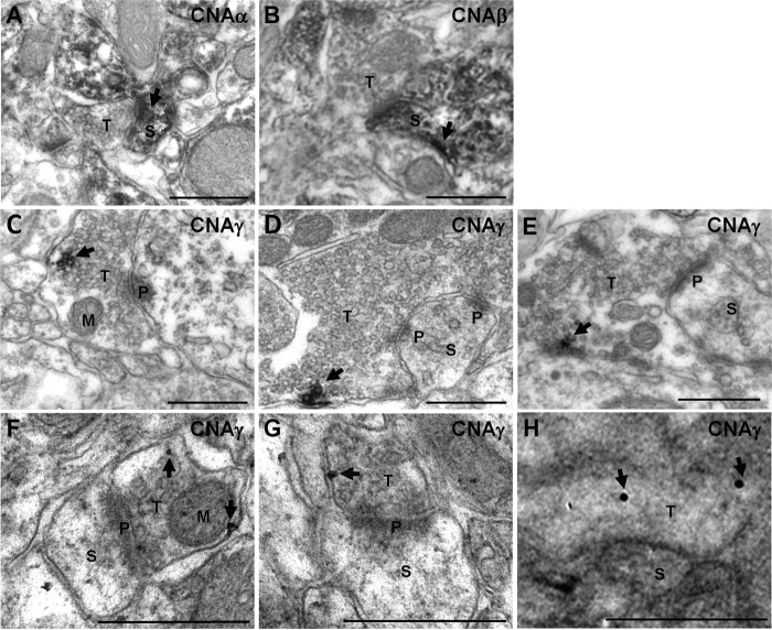
FIGURE 3.
CNAγ is present in presynaptic terminals in the hippocampus. A—E, pre-embedding immuno-EM micrographs were performed on mouse hippocampus sections using anti-mouse CNAα (A), CNAβ (B), or CNAγ (C–E) antisera. F—H, post-embedding immunogold micrographs on mouse hippocampus sections using anti-mouse CNAγ antisera. Arrows indicate CNA labeling. Scale bars = 500 nm. S, spine; T, presynaptic terminal; P, postsynaptic density; M, mitochondria.

