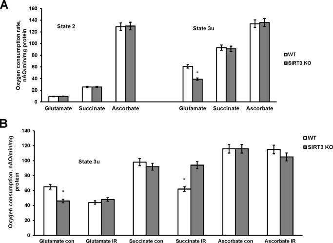
FIGURE 6.
Respiratory chain activity in baseline and ischemic cerebral mitochondria from WT and Sirt3 KO mice. Mitochondria were purified from WT and Sirt3 KO mouse brain at baseline (A) and from the WT and Sirt3 KO mouse brain hemispheres ipsilateral (IR) and contralateral (con) to damage following 1 h of MCAO/24 h of reperfusion (B). Mitochondrial respiration was measured by recording oxygen consumption with a Clark-type oxygen electrode in the presence of complex I substrate 5 mm glutamate plus 5 mm malate (glutamate), complex II substrate 10 mm succinate (succinate), and complex IV substrate 1 mm ascorbate plus 250 μm N,N,N′,N′-tetramethyl-p-phenylenediamine (ascorbate) without (state 2) or with 50 μm 2,4-dinitrophenol (state 3u). Data are means ± S.E., *, p < 0.05, n = 16.

