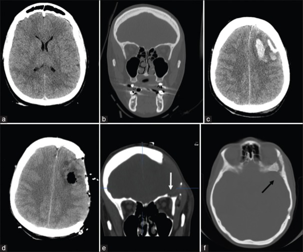
Figure 1.
(a and b) The immediate postinjury thin-slice computed tomography scan with coronal reconstruction showed generalized brain edema without left orbital roof fractures. (c) Computed tomography scan 24 h after trauma revealed a left-sided frontal contusion causing midline shift and increased diffuse brain swelling. (d and e) Postoperative computed tomography scan showing left decompressive craniotomy and evacuation of frontal contusion with a small bony defect in the roof of the orbit (see arrow) probably related to an unintentional opening of the orbit during keyhole burr hole without evidence of encephalocele. (f) Computed tomography scan after cranioplasty confirming the small bone opening on the orbital roof (see arrow)

