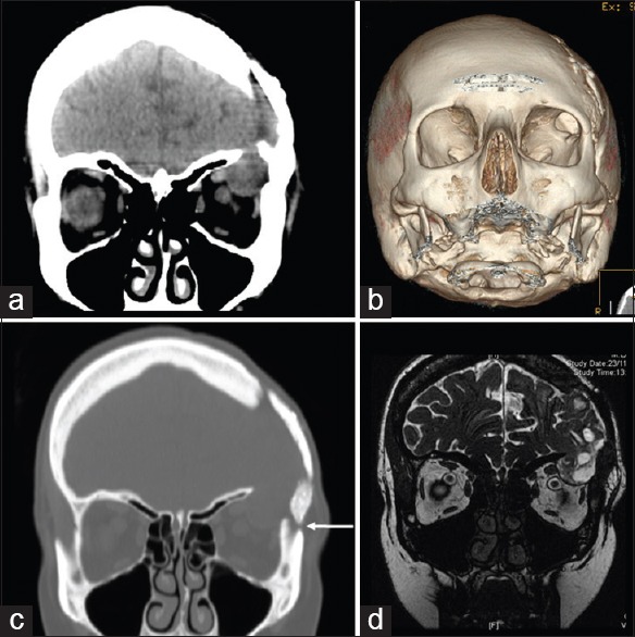
Figure 2.

(a-c) Thin-slice orbital computed tomography sections with three-dimensional and coronal reconstruction (2 years after the cranioplasty) revealed the enlargement of the lateral orbital roof bony defect associated with intraorbital encephalocele. The arrow indicates the burr hole, filled by autologous bone dust mixed with a bone substitute, too close to the orbital wall. (d) Magnetic resonance imaging image confirming the herniation of brain matter into the left orbit
