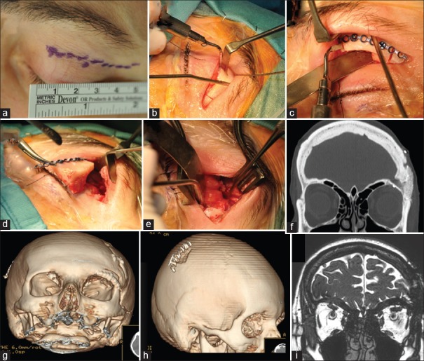
Figure 3.
Abcdefghi: Intraoperative images and postoperative thin-slice computed tomography scan with three-dimensional reconstruction. (a) Left superior blepharoplasty incision. (b and c) Supra-transorbital keyhole approach with the combination of an orbital osteotomy with a supraorbital minicraniotomy. The use of piezoelectric scalpel allows to realize precise and thin osteotomies, for better future bone healing and better aesthetic result. (c and d) The one-piece bone flap includes the frontal bone, and the orbital rim using the defect of the orbital roof as the posterior edge of the orbitotomy. The one-piece bone flap was then fixed by titanium low-profile miniplate and screws. (e) Intraoperative working area with the exposure of both orbital and intracranial compartments. (f-i) Postoperative thin-slice computed tomography scan with three-dimensional reconstruction and magnetic resonance imaging showing left orbital roof reconstruction with autologous bone, obtained from the split calvarial parietal bone contralateral to the cranioplasty

