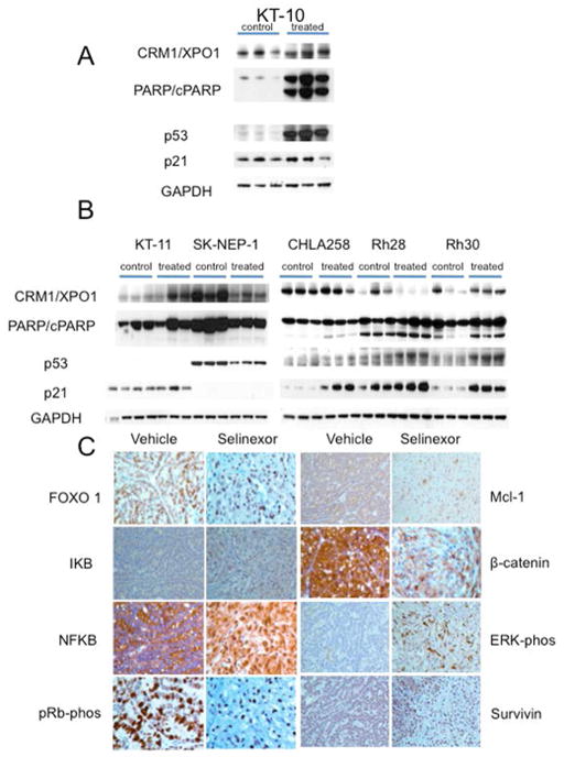
Figure 4.
A. Pharmacodynamic changes induced by selinexor. A. KT-10 Wilms tumor. Tissue was harvested 24 hr after a single dose of selinexor (10 mg/kg); B. Tumors were harvested 2 hr after dose six of selinexor (10 mg/kg/dose). With the exception of SK-NEP1, all tumors are p53 wild type by sequence analysis. Three control and 3 tumors from treated mice were used for each xenograft line. GAPDH was used as a loading control. B. In vivo treatment with selinexor blocked XPO1 cargos in the nucleus and reduced the expression of signaling proteins that are associated with cell proliferation. Sections from vehicle and selinexor treated KT-10 were analyzed by immunohistochemistry (IHC). Cells from tumors that were treated with selinexor show increased nuclear accumulation of FOXO1, IKB, NFKB, pRb, ERK and Survivin. In addition the IHC slides show reduction in Mcl-1 and β-catenin.

