Fig. 1.

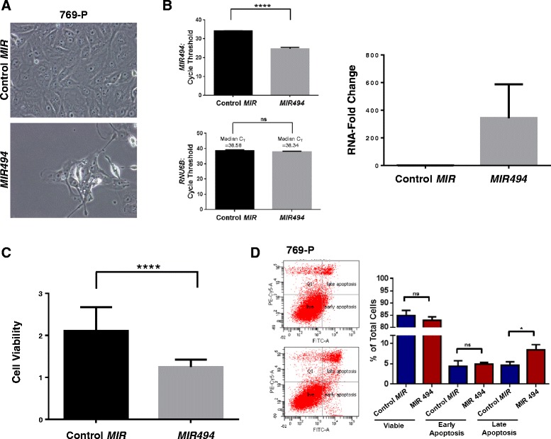
MIR494 modulates cell viability by altering the apoptotic response and LC3B levels. a Light microscope images of 769-P cells expressing MIR494 or control MIR were captured 96 h post-transfection. Representative images at 40× magnification are presented. b miRNA isolation and quantification of MIR494 was performed in 769-P cells expressing control or MIR494. Cycle threshold changes (left panel) and RNA-fold changes (right panel) are presented. Three independent experiments were performed. c 769-P cells expressing MIR494 or control MIR were re-seeded into 96-well plates; following 96 h post-transfection, cell viability was assessed. A total of five independent replicates were performed. d Annexin V-PI staining was performed in 769-P cells expressing MIR494 or control MIR at 96 h post-transfection. Raw data plots are shown as log fluorescence values of annexin V-FITC and PI on the X and Y axis, respectively. The percentage of viable, early apoptotic, and late apoptotic cells are shown. Three independent replicates were performed
