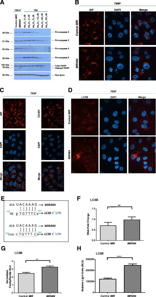
Fig. 3.

MIR494 induces LC3B mRNA expression and LC3B-associated punctae. a T80 cells were seeded at 250,000 cells/well. Twenty-four hours post-seeding, cells were treated with the indicated doses of As2O3 for 18 h, after which protein lysates were collected. Samples were run on a SDS-PAGE gel and analyzed via western blotting using the indicated antibodies. Two independent experiments were performed. b Indirect immunofluorescence was performed on 769-P cells transfected with MIR494 or control MIR at 96 h post-transfection for AIF. Three independent experiments were performed. Representative images are presented. c Indirect immunofluorescence was performed on 769-P cells for AIF or COXIV. Representative images are presented. d 769-P cells expressing MIR494 were subjected to immunofluorescence staining for LC3B. Two independent experiments were performed. Representative images are presented. e The schematic depicts MIR494 binding sites in the 3′-UTR of LC3B (2 imperfect binding sites). Grey boxes indicate the binding region on the mRNA transcript of LC3B. f Total RNA was isolated from 769-P cells expressing MIR494 or control MIR and used for real-time PCR. Relative RNA-fold changes are presented for LC3B. Three independent experiments were performed. g T80 cells were transfected with pEZX-MT01 plasmid harboring the 3′-UTR of LC3B downstream of the luciferase gene in the absence or presence of MIR494. Three independent experiments were performed. h T80 cells were transfected with pLightSwitch plasmid harboring the promoter of LC3B upstream of the luciferase gene in the absence or presence of MIR494. Three independent experiments were performed
