Fig. 5.

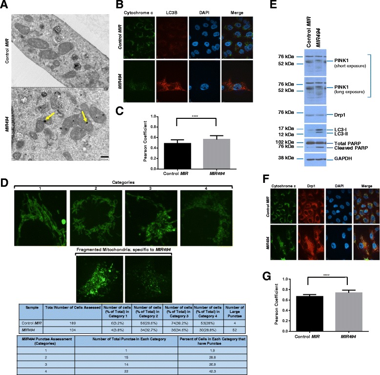
MIR494 expression leads to mitochondrial changes. a TEM images were captured from control or MIR494 transfected 769-P cells. Yellow arrowheads indicate electron dense regions in the mitochondria. b Indirect immunofluorescence was performed on 769-P cells transfected with MIR494 or control MIR at 96 h post–transfection for LC3B and cytochrome c. Three independent experiments were performed. Representative images are presented. c Pearson coefficients are shown for data presented in (b). d Mitochondrial patterns were segregated into four distinct categories as described in Results. The quantified data is presented in tabular form. e Protein lysates collected from MIR494 or control MIR expressing 769-P cells were re-run on a SDS-PAGE gel and analyzed via western blotting using the indicated antibodies. Three independent experiments were performed. f Indirect immunofluorescence was performed on 769-P cells transfected with MIR494 or control MIR at 96 h post-transfection for Drp1 and cytochrome c. Three independent experiments were performed. Representative images are presented. g Pearson coefficients are shown for data presented in (f)
