
Fig. 1.
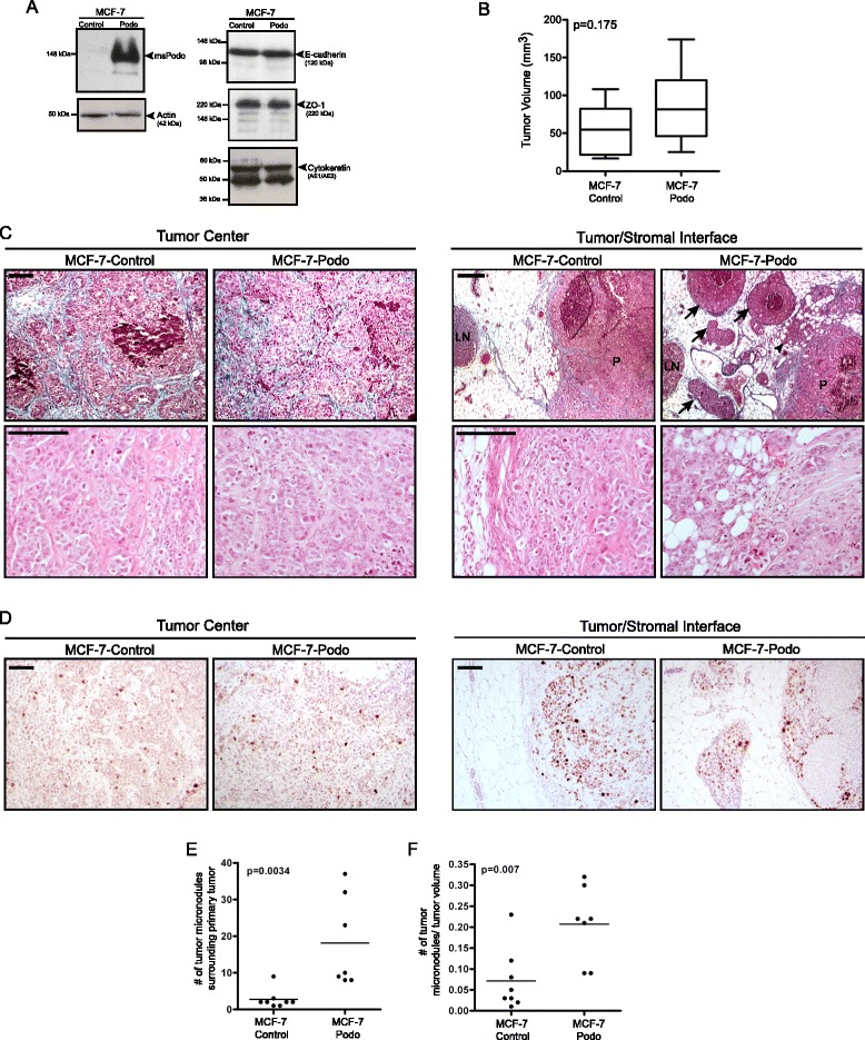
Podocalyxin overexpression induces collective tumor invasion in vivo. a Western blot analyses of whole cell lysates show that total expression of E-cadherin, the tight junction protein ZO-1, and epithelial cytokeratins were unaffected by stable podocalyxin overexpression in MCF-7-podo cells compared with MCF-7-control cells. b Podocalyxin overexpression did not significantly affect overall estrogen-dependent MCF-7 cell-derived tumor growth after orthotopic xenotransplantation (n = 8 for each condition; tumors were excised after 6 weeks; p = 0.175, two-tailed, unpaired Student’s t test). c Representative images of trichrome-stained tumor sections (upper panels) show that MCF-7-control and MCF-7-podo xenografts both contained densely packed tumor cells at the center of the primary tumor nodule. Both tumor types also had small areas of visible necrosis and developed a collagenous stroma (light blue staining) that surrounded nests of cohesive tumor cells. Representative images of the tumor/host stromal interfaces show that, compared with controls, MCF-7-podo cells more extensively protruded into the surrounding stroma at the edge of tumor (arrowhead). These tumors also formed multiple small cohesive tumor foci, or micronodular bud-like structures (arrows), that appeared to be separate from the primary tumor nodule (labeled P; local lymph node is labeled LN). Higher power H & E images (lower panels) indicate that there was no a difference in tumor cell density within either the centers or edges of the lesions at the tumor/stromal interface. Scale = 100 μm. d Tumors were immunostained with the proliferation marker Ki-67 (brown). While there was an overall increase in Ki-67-positive cells at the tumor/stromal interface, there was no discernible difference in Ki-67 frequency between MCF-7-control and MCF-7-podo cells either at the center of the tumors (left panel) or at the tumor/stromal interface (right panel). Scale = 100 μm. e, f The number of observable micronodules was quantified either per tumor e or per tumor volume f. Note that, in both cases, there was a statistically significant increase in the MCF-7-podo tumors compared with the control tumors (two-tailed, unpaired Student’s t test)