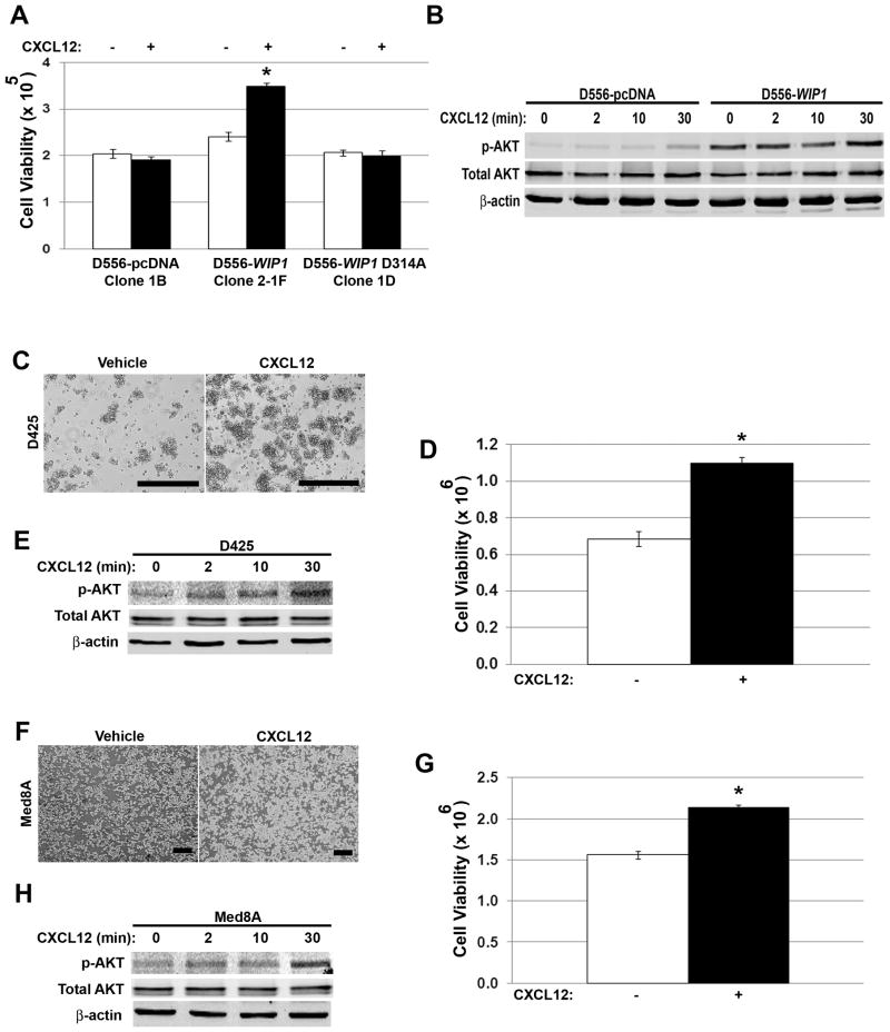
Figure 3. Stimulation with SDF1α activates PI-3 kinase signaling and promotes growth of WIP1 high-expressing medulloblastoma cells.
(A) Number of viable D556 stably-transfected cells by trypan blue exclusion (i.e. viability; Y-axis), 48 hours following serum starvation and stimulation with vehicle (-) or CXCL12 (+; SDF1α, 1 μg/mL). *, p < 0.0002. (B) Western blotting of whole cell lysates from (A) for Ser473-phosphorylated and total AKT. (C, F) Photomicrographs and (D, G) number of viable D425 and Med8A cells by trypan blue exclusion, respectively, 48 hours following serum starvation and SDF1α stimulation, as above. Scale bars, 400 μm. *, p < 0.0002. (E, H) Western blotting of whole cell lysates from (D, G) for serine 473-phosphorylated and total AKT. Error bars, standard deviation. Experiments were repeated at least three times.

