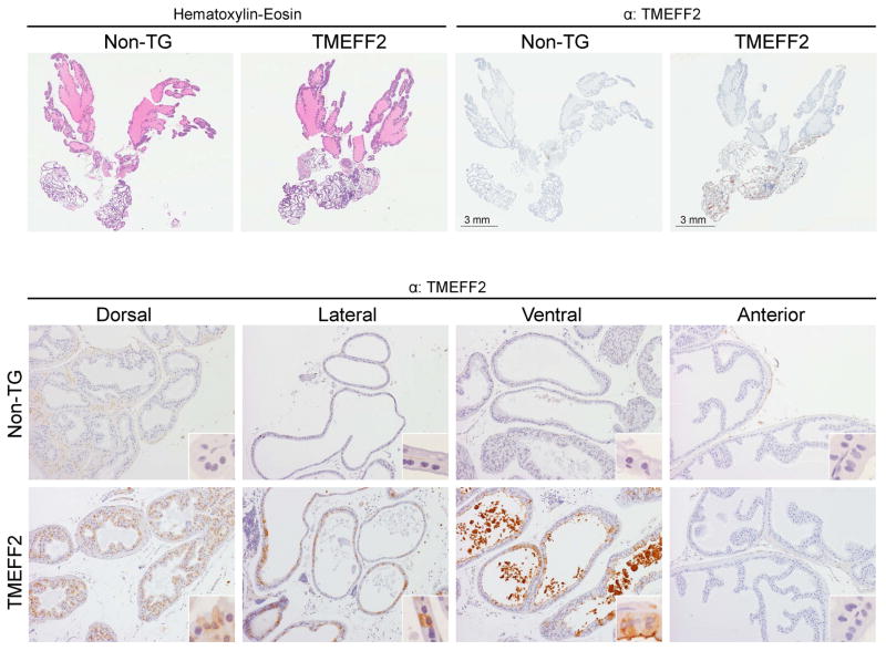
Figure 3. Histological and immunohistochemical analysis of the transgenic TMEFF2 mouse.
Top: Representative images of whole prostates stained with H&E (left) or IHC with an antibody against TMEFF2 (top right). Bottom: Representative IHC images of the different prostatic lobes from non-transgenic or transgenic-TMEFF2 mice stained with an antibody against TMEFF2. Original magnification is 10x and in inserts is 60x.

