
Fig. 8.
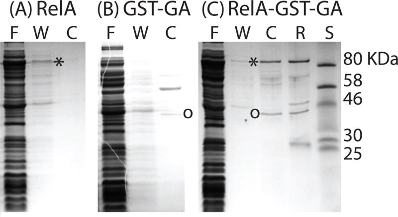
ComGA binds to RelA from B. subtilis (RelABsu). Three E. coli strains, expressing RelABsu (A), GST-ComGA (B) and both proteins (C) were grown, lysed and the extracts were passed over a glutathione column. Proteins were eluted following on-column cleavage with PreScission protease and the various fractions were analyzed by SDS-PAGE. “F”, “W” and “C” refer to flow through, wash and cleaved fractions respectively. “R” refers to the material remaining on the resin and the lane labeled “S” contains molecular weight standards. Asterisks and circles indicate the locations of RelABsu and of ComGA respectively. The identity of RelABsu was confirmed by mass spectrometry.