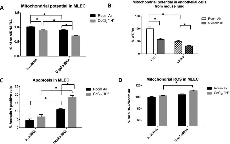
Figure 4.
Mouse lung endothelial cells (MLEC) after Ucp2 silencing and kept at room air or challenged with CoCl2 “IH”. Scrambled siRNA (Sc) was used as negative control. A. Mitochondrial membrane potential as assessed by FACS in room air or after CoCl2 “IH”. B. Mitochondrial membrane potential as assessed by FACS in EC isolated from Flox and VE-KO lungs in room air and after 5 weeks of IH. C. Apoptosis as assessed by annexin-PI FACS in room air or after CoCl2 “IH”. D. Mitochondrial ROS production as assessed by FACS in room air or after CoCl2 “IH”. *p<0.05.

