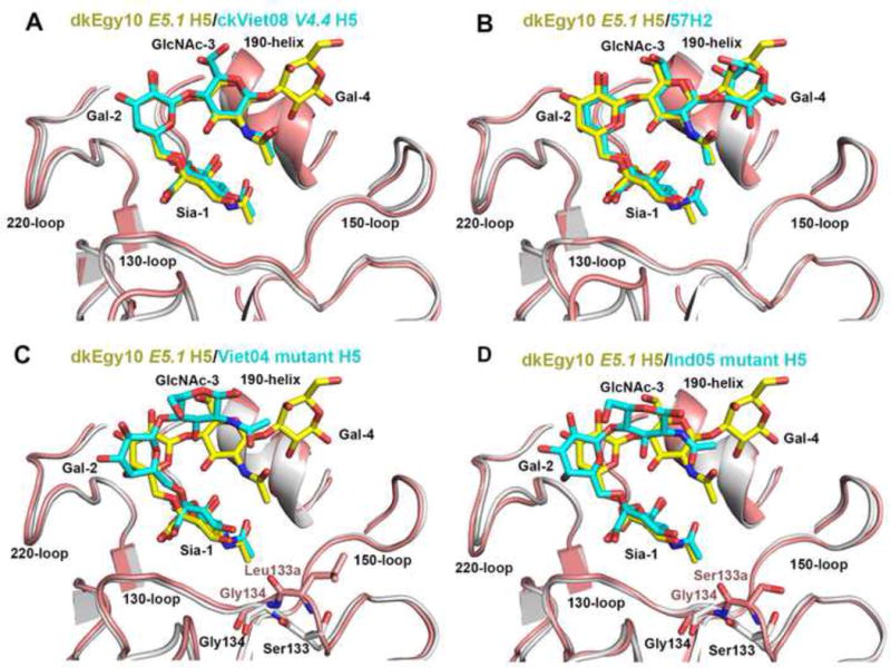
Figure 3. Structural comparison of LSTc bound to E5.1 H5 HA with other HAs.

The receptor binding subdomain (residues 117–265) was used to superimpose HA structures. (A) Superimposition of the RBS of E5.1 HA (grey) with LSTc (yellow carbon atoms) and the RBS of V4.4 H5 HA (pink) with LSTc (cyan carbon atoms). The same coloring scheme is used in (B), (C) and (D). (B) Superimposition of the RBS of E5.1 HA with LSTc and human H2 HA with LSTc (PDB code 2WR7). (C) Superimposition of the RBS of E5.1 HA with LSTc and Viet04 mutant H5 HA with LSTc (PDB code 4KDO). The side chain of Leu133a insert of Viet04 mutant HA is shown in pink carbon atoms. (D) Superimposition of the RBS of E5.1 HA with LSTc and Ind05 mutant H5 HA with LSTc (PDB code 4K67). The side chain of Ser133a insert of Ind05 mutant HA is shown in pink carbon atoms. Figures (A), (B), (C) and (D) are in the same orientation.
