
Figure 6.
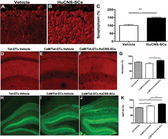
HuCNS‐SC transplantation increases presynaptic markers and axonal sprouting. (A) Immunolabeling of synaptophysin in vehicle injected 3xTg‐AD mice. (B) HuCNS‐SC injection results in a 47% increase in synaptophysin levels in the stratum radiatum of CA1, quantified in (C, N = 5, t‐test P < 0.008). (D) Synapsin in control unlesioned mice reveals a typical pattern of presynaptic innervation within the stratum radiatum of CA1. (E) Surprisingly, synapsin levels are only slightly diminished in lesioned mice. (F) In contrast, HuCNS‐SC transplantation significantly increases the density of presynaptic innervation, quantified in (G, N = 8–12, ANOVA P < 0.05, Fishers PLSD P = 0.016). (H) Compared to control unlesioned mice, we also detected a significant increase in GAP‐43 immunoreactivity within the dentate gyrus of lesioned CaM/Tet‐DTA mice with vehicle injections (I). More importantly, we observed a further enhancement of GAP‐43 expression in lesioned mice transplanted with HuCNS‐SC (J), quantified in (K). (N = 10, ANOVA P < 0.05, Fishers PLSD P = 0.028, P = 0.006, P = 0.0001). Data presented as mean ± SEM. [Color figure can be viewed in the online issue, which is available at wileyonlinelibrary.com.]