
Fig. 4.
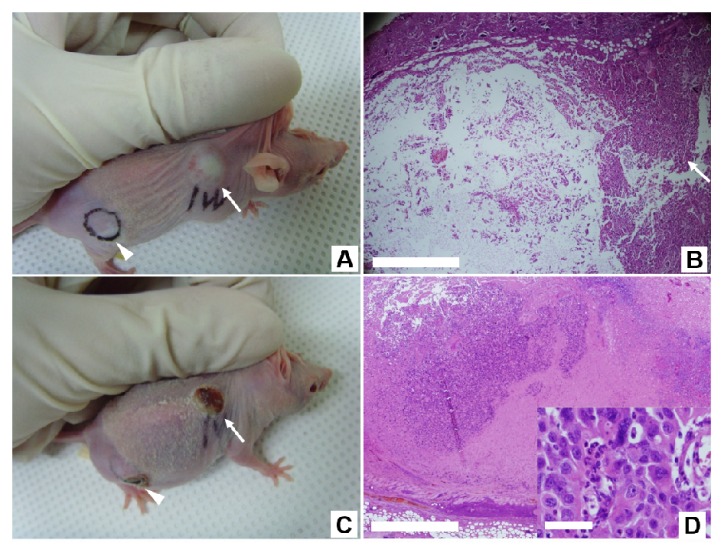
The effect of PTT on the control and intratumoral injection groups. The xenograft tumor mass was illuminated using a NIR laser at an irradiance of 1 W/cm2 for 2 minutes without (A and B) and with (C, D, E and F) the injection of NS-loaded macrophages. (A) A photograph of a mouse with xenograft tumor (marked as a violet dot) where NS-loaded macrophages were not injected. (B) The H&E stained image of a tissue section from the tumor site after the laser irradiation. The tumor was not affected by laser irradiation as the zoom-in image (inset in Fig. (B)) of the tumor area (marked as a red box) shows that the cellular structures were well preserved. On the other hand, the skin and subcutaneous tissues indicated as a blue box were detached from deep tissue around the tumor because of the edematous change resulting from the heat. (C) A photograph of a mouse with xenograft tumor (white arrow) to which NS-loaded macrophages were injected. (D) The H&E image of a tissue section from the tumor site immediately after administrating PTT. Extensive cellular destruction and dead spaces are evident on the left side of tumor interior, but the other portion shows intact tumor cells (white arrow). (E) A photograph of the same mouse in (C) taken after two weeks of the PTT. Larger and deeper crust formation occurred on the tumor site (white arrow). (F) The H&E stained image of a tissue section from the mouse in Fig. E. The degraded portion within the tumor displays acellular fibrotic tissue with dead cells. Live tumor cells were identified near the destructed portion. Scale bars in A, C and E corresponds to 1 mm, those in B, D and F are 500 μm, and the scale bars in their insets are 50 μm.