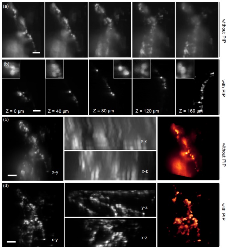
Fig. 6.
Visualization of 3D-cultured cell branches using PIP-integrated conventional microscope. (a) Sequential image slices acquired by a conventional inverted microscope using 10X/0.3 air objective. Out-of-focus excitation caused drastic image deterioration. (b) Sequential image slices acquired at the same z depths using identical wide-field detection plus 10 μm optical sectioning of PIP. (c) - (d) The maximum intensity projections (MIPs) and the volume renderings obtained without and with PIP, respectively. y-z, x-z projections of the 3D reconstructed images were also shown. The incorporation of PIP achieved significant axial resolution enhancement and background noise reduction. The cells geometry and intracellular morphology were also exclusively revealed by clear volume rendering in PIP group (rightmost). Scale bars in all images are 100 μm.

