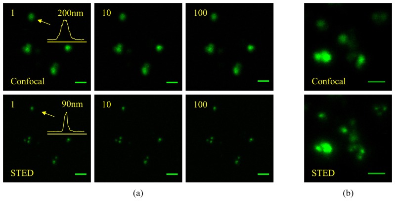
Fig. 4.
(a) Consecutive confocal and continuous STED imaging of 70 nm sized nanodiamonds immobilized on a glass slide. The increase in resolution leads to reveal details blurred in the conventional confocal images. The inset on the first images is the profile along the particle indicated with an arrow. It allows for the estimation of the resolution given by STED to be at least 70 nm for a particle bigger than 50 nm (cf. Appendix B). The multiple scans illustrate the perfect photostability. Even if for the STED scan images, high depletion intensity was used (I = 130 MW/cm2, 256 × 256 pixels with 3 ms dwell time), no change in the recorded signal level is observed after 100 scans. (b) Confocal and STED image revealing the inhomogeneity of the nanoparticles and their tendency to aggregate. Scale bars are 500 nm.

