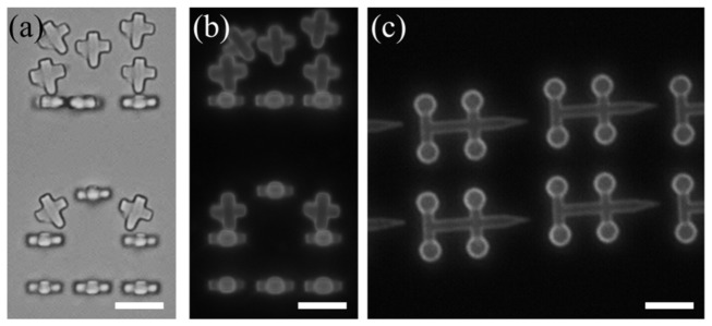
Fig. 3.
Optical microscopy images of polymerized structures made for indirect cell trapping. Brightfield (a) and fluorescence (b) microscopy images of crosses and fluorescence image of four-spheroids manipulators (c) coated with fluorescent streptavidin. The structures are still attached onto their substrates, except some of the crosses. Scale bar: 10 μm.

