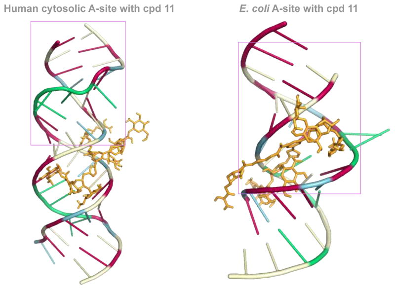
Fig. 5.
Docked structures of the L-arginine-linked NEO dimer 11 (depicted as orange stick) with the E. coli (right) and human RNA (left) A-sites. The nucleoside bases are colored as follow: A = green, G = red, C = pale yellow, and U = blue. The pink box represent the residues that are identical to those presented in Fig. 2.

