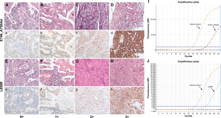
Figure 1.
Representative pictures for IHC staining and ARMS analysis on mutant EGFRs.
Notes: H&E staining (A–H) and immunohistochemical staining (a–h) for EGFR E746_A750del (A–D for H&E staining, a–d for match IHC staining) and L858R (E–H for H&E staining, e–h for match IHC staining) mutation-specific antibody in tumor samples from patients with primary lung adenocarcinomas, respectively. (A and a, E and e) No IHC staining in malignant glandular epithelium for EGFR mutant protein (0+); (B and b, F and f) Faint IHC membrane staining in. 10% tumor cells (1+); (C and c, G and g) Moderate complete IHC membrane staining in. 10% tumor cells (2+); (D and d, H and h) Strong complete IHC membrane staining in. 10% tumor cells (3+) (magnification, 200×). Representative results for exon 19 E746_A750del (I) and exon 21 L858R (J) mutations by ARMS method are also shown.
Abbreviations: H&E, hematoxylin–eosin; EGFR, epidermal growth factor receptor; IHC, immunohistochemistry; ARMS, amplification-refractory mutation system.

