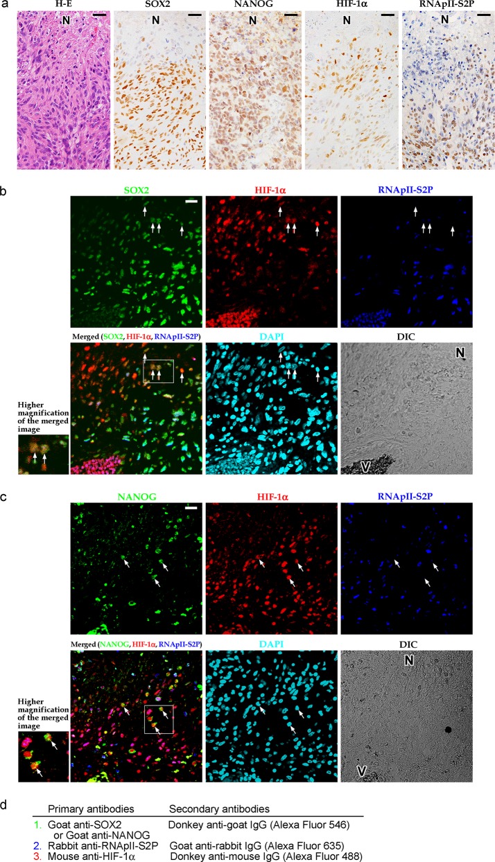
Fig 1. Tumor cells with a SOX2+ (or NANOG+) HIF-1α+ RNApII-S2P-/low phenotype are found in human glioblastoma tissues.
a: Hematoxylin-eosin (H-E) staining and single-color immunostaining for SOX2, NANOG, HIF-1α, and RNApII-S2P (serine 2 phosphorylation of the C-terminal domain of RNA polymerase II) in tumor tissue from a representative case of glioblastoma. Suppression of RNApII-S2P immunoreactivity was noted around the necrotic area (N). b: Triple immunofluorescent staining for SOX2, HIF-1α, and RNApII-S2P. SOX2+ HIF-1α+ RNApII-S2P-/low cells (arrows) were found around the necrotic area (N). c: Triple immunofluorescent staining for NANOG, HIF-1α, and RNApII-S2P. NANOG+ HIF-1α+ RNApII-S2P-/low cells (arrows) were found around the necrotic area (N). Insets at lower left of the panels b and c show higher magnification of the boxed areas in the merged images. N, necrotic area; V, blood vessels; DAPI, 4′,6-diamidino-2-phenylindole (nuclear stain); DIC, differential interference contrast image. Scale bars, 25 μm. d: Summary of the staining methods used in b and c. Sections were incubated with goat anti-SOX2 (b) or anti-NANOG (c) antibody and then with Alexa Fluor 546-conjugated donkey anti-goat IgG secondary antibody. Next, rabbit anti-RNApII-S2P antibody and then Alexa Fluor 635-conjugated goat anti-rabbit IgG secondary antibody were applied. The sections were reacted with mouse anti-HIF-1α antibody and then with biotinylated donkey anti-mouse IgG secondary antibody and Alexa Fluor 488-conjugated streptavidin.

