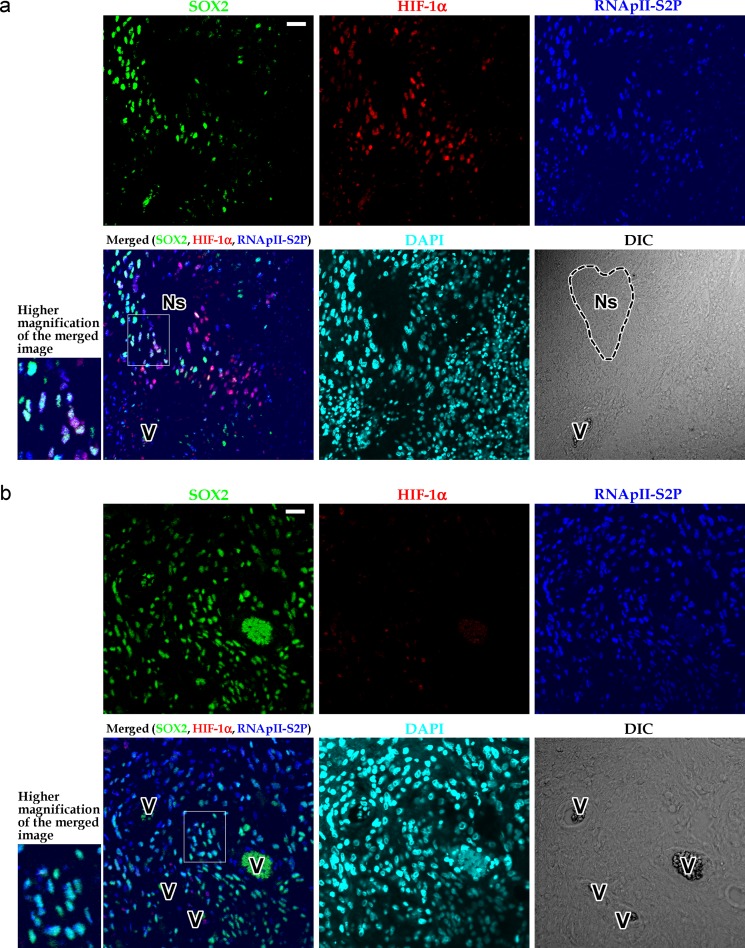
Fig 4. Triple immunofluorescent staining for SOX2, HIF-1α, and RNApII-S2P in zones around small pseudopalisading necroses or in areas showing no necrotic changes.
a: Glioblastoma tissue containing small pseudopalisading necrosis. Although SOX2+ and/or HIF-1α+ tumor cells were found, they were RNApII-S2P+. Therefore, SOX2+ HIF-1α+ RNApII-S2P-/low cells were not observed. b: Glioblastoma tissue showing no necrotic changes. SOX2+ HIF-1α+ RNApII-S2P-/low cells were not found. Insets at lower left of the panels a and b show higher magnification of the boxed areas in the merged images. Ns, small pseudopalisading necrosis; V, blood vessels; DIC, differential interference contrast image. Scale bars, 25 μm.

