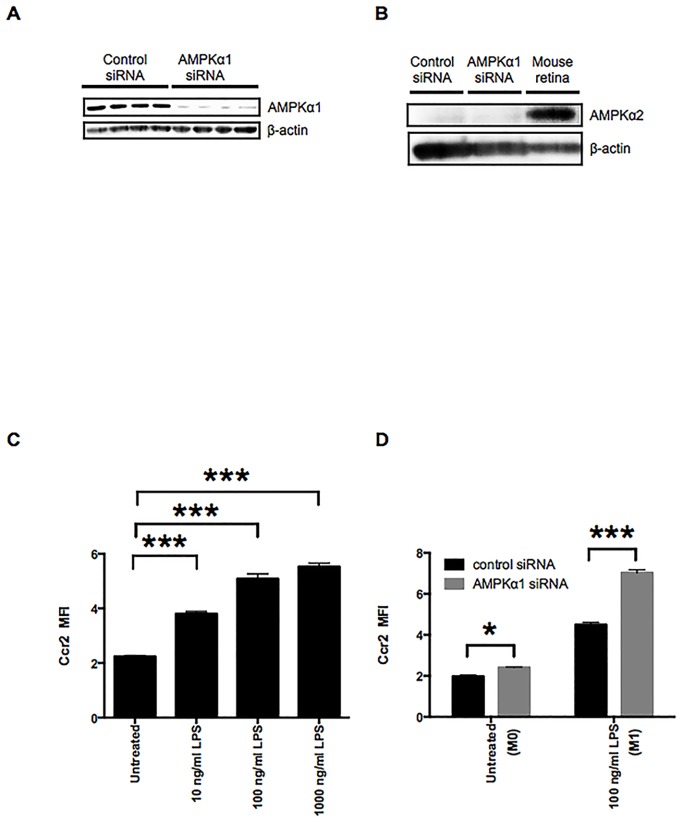
Fig 1. AMPKα1 negatively regulates Ccr2 expression in the M0 and the LPS-stimulated M1 macrophages.
A: Reduced AMPKα1 protein levels in macrophages treated with AMPKα1 siRNA RAW264.7 were confirmed by Western blotting of whole cell lysates. β-actin was probed as an internal control. B: Whole cell lysates of RAW264.7 macrophages treated with either control or AMPKα1 siRNA were examined by Western blotting to confirm compensation by AMPKα2. Tissue lysates prepared from mouse retina were used as a positive control for expression of AMPKα2 protein. β-actin was probed as an internal control. C: Ccr2 expression on RAW264.7 macrophages was analyzed by flow cytometry. RAW264.7 macrophages were stimulated with 10–1000 ng/ml of LPS for 12 h. D: Flow cytometry analysis of Ccr2 expression on RAW264.7 macrophages treated with either control or AMPKα1 siRNA. RAW264.7 macrophages were stimulated with 100 ng/ml of LPS for 12 h to induce the M1 state. n = 3. ***, p < 0.001.

