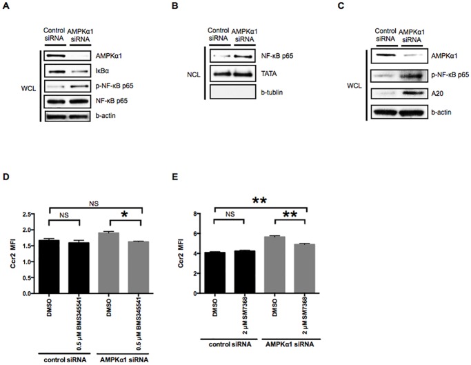
Fig 3. Activation of the NF-κB pathway reverses AMPKα1-dependent downregulation of Ccr2 expression in M0 macrophages.
A: Whole cell lysates (WCL) of RAW264.7 macrophages treated with either control or AMPKα1 siRNA were examined by Western blotting to determine IκBα degradation and NF-κB p65 phosphorylation. β-actin was probed as an internal control. B: Nuclear cell lysates (NCL) of RAW264.7 macrophages treated with either control or AMPKα1 siRNA were examined by Western blotting to determine activation of a NF-κB pathway. TATA and β-tubulin antibodies were used to confirm equal protein loading and to assess the relative purity of the nuclear cell lysates. C: Whole cell lysates (WCL) of RAW264.7 macrophages treated with either control or AMPKα1 siRNA were examined by Western blotting to determine the expression of A20. β-actin was probed as an internal control. D and E: RAW264.7 macrophages treated with either control or AMPKα1 siRNA were additionally treated with IKK inhibitor (BMS345541, 0.5 μM) and NF-κB inhibitor (SM7368, 2 μM) for 12 h. Ccr2 expression was analyzed by flow cytometry. n = 3. *, p < 0.05; **, p < 0.01.

