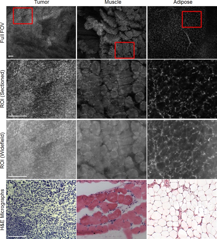
Fig 4. Site-level images.
Representative SIM images of three different tissue types commonly found in margin images: tumor, muscle, and adipose tissues. For the smaller ROIs, both the sectioned (structured illumination) and widefield (standard illumination) images are shown to demonstrate the enhanced contrast that SIM provides. Examples of H&E micrographs are also shown, but it should be noted the H&E is not taken from the exact site of the corresponding SIM images because specific site level fiducial markers were not used in this study. Scale bars are 200 μm.

