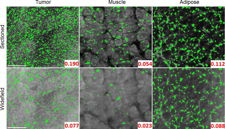
Fig 8. Segmented tissue images using the finalized MSER parameters.
Application of the finalized set of MSER parameters to both the uniform and sectioned representative image set. It is evident that the sectioned images provide a clear benefit with contrast enhancement which assists the MSER algorithm in accurately identifying nuclei. The finalized set of MSER parameters used for these segmentation results were MinArea = 3, MaxArea = 15, MinDiversity = 0.5, MaxVariation = 2.5, and Delta = 10. The segmented area fraction is displayed in the bottom right of each image. It is apparent that a lower segmented area is seen in widefield illumination images, due to the decreased contrast. Scale bars are 200 μm.

