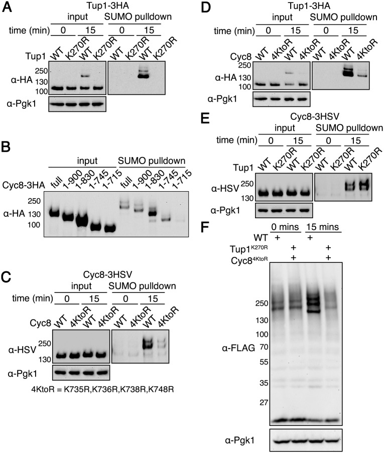
Fig 4. Identification of Tup1 and Cyc8 sumoylation sites.
(A) His6-FLAG-SMT3 cells expressing either wild-type Tup1-3HA or Tup1K270R-3HA from the endogenous TUP1 promoter were examined for Tup1 sumoylation at 0 or 15 minutes of hyperosmotic stress (1.2M sorbitol) as in (Fig 2D). (B) His6-FLAG-SMT3 cells expressing the indicated Cyc8-3HSV deletion mutant were subject to hyperosmotic stress (1.2M sorbitol) for 15 minutes. Cell lysates were generated and subject to the same analysis as in (Fig 2D). (C) His6-FLAG-SMT3 cells expressing either wild-type Cyc8-3HSV or Cyc84KtoR-3HSV from the endogenous CYC8 promoter were examined for Cyc8 sumoylation as in (Fig 2D). (D) His6-FLAG-SMT3 cells expressing wild-type Tup1-3HA from the endogenous TUP1 promoter and either wild-type Cyc8-3HSV or Cyc84KtoR-3HSV from the endogenous CYC8 promoter were examined for Tup1 sumoylation as in (A). (E) His6-FLAG-SMT3 cells expressing wild-type Cyc8-3HSV from the endogenous CYC8 promoter and either wild-type Tup1-3HA or Tup1K270R-3HA from the endogenous TUP1 promoter were examined for Cyc8 sumoylation as in (C). (F) 6His-FLAG-SMT3 wild-type or tup1K270Rcyc84KtoR cells were subjected to hyperosmotic stress (1.2M sorbitol) for 0 or 15 minutes, and global sumoylation patterns examined by western analysis using an anti-FLAG antibody.

