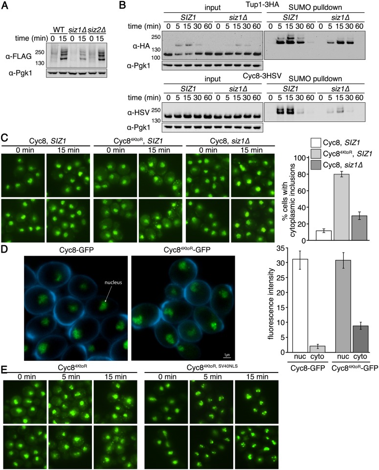
Fig 7. Siz1 is a SUMO ligase that influences Cyc8 sumoylation.
(A) His6-FLAG-SMT3 wild-type, siz1Δ, or siz2Δ cells were subject to hyperosmotic stress (1.2M sorbitol) for 0 or 15 minutes. Global sumoylation patterns were examined by western analysis using an anti-FLAG antibody. (B) His6-FLAG-SMT3 wild-type or siz1Δ cells expressing either Tup1-3HA or Cyc8-3HSV from their endogenous promoters were subject to hyperosmotic stress (1.2M sorbitol) over a 60-minute time course. Cell lysates were generated and subject to metal affinity chromatography to purify sumoylated proteins. Cell lysates (input) and purified sumoylated proteins (SUMO pulldown) were subject to SDS-PAGE and western analyses using anti-HA (Tup1) or anti-HSV (Cyc8) antibodies. (C) SIZ1 or siz1Δ cells expressing wild-type Cyc8 or sumoylation-deficient Cyc84KtoR tagged with GFP from the endogenous CYC8 promoter were exposed to hyperosmotic stress (1.2M sorbitol) for 0 and 15 minutes, fixed at the times indicated, and imaged by fluorescence microscopy. Six fields of cells for each condition, with ≥40 cells/field, were counted for the presence of cytoplasmic inclusions. Data represent the average percentage of cells that contained cytoplasmic inclusions within the six fields. Error bars are the standard deviation. (D) Confocal 3D images and quantitation of Cyc8-GFP and Cyc84KtoR-GFP localization in the nucleus and cytoplasm. The nuclear and cytoplasmic fluorescence intensities were measured using NIS-Elements software (Nikon). Data represents the average of at least 100 cells in at least 10 fields in 3 independent experiments. Error bars represent the standard deviation. (E) Cells expressing GFP-tagged versions of Cyc84KtoR or Cyc84KtoR with the SV40 NLS appended to the C-terminus (both expressed from the endogenous CYC8 promoter) were exposed to hyperosmotic stress (1.2M sorbitol) for 0, 5, and 15 minutes, fixed at the times indicated, and imaged by fluorescence microscopy.

