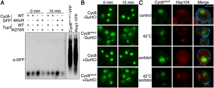
Fig 11. Cyc8 inclusions are not SDS-resistant, Hsp104-dependent amyloids.
(A) Semi-denaturing detergent agarose gel electrophoresis (SDD-AGE) of lysates from the strains used in Fig 6A. Cells were exposed to 0 or 15 minutes of hyperosmotic stress (1.2M sorbitol), and lysates were mixed with semi-denaturing buffer before loading into the agarose gel. Lysates from strains overexpressing either Rnq1-YFP or Cyc8441-677-YFP are also included as positive controls for SDS-resistant amyloid formation. Western analysis was performed with anti-GFP antibody. The two images are different exposures of the same blot. (B) Fluorescence microscopy of cells expressing GFP-tagged wild-type or sumoylation-deficient Cyc8 after 3 passages on either YPD or YPD+4mM guanidinium hydrochloride (GuHCl). Cells were exposed to 0 or 15 minutes of hyperosmotic stress (1.2M sorbitol), fixed, and imaged by fluorescence microscopy. All constructs were integrated at the gene’s endogenous locus and expressed from the endogenous promoter. (C) Confocal 3D images of cells expressing mutant Cyc84KtoR tagged with GFP and Hsp104 tagged with mCherry were subject to a 15 minute 42°C heat shock to induce Hsp104 inclusions, 15 minute hyperosmotic stress (1.2M sorbitol) to induce Cyc84KtoR inclusions, or both. Cell wall was visualized by staining with Calcofluor White.

