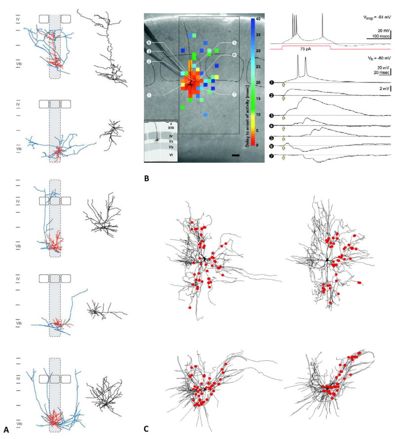
Figure 2. Examples of primary discoveries made with digital reconstructions.
A) Diversity of excitatory neurons in layer 6b of the rat barrel cortex from the Feldmeyer archive of NeuroMorpho.Org (adapted from Marx and Feldmeyer 2013). B) Morphological and electrophysiological characterization of newly identified pyramidal neurons from the Staiger archive (adapted from Schubert and others 2006). The left photomicrograph of the native coronal slice is superimposed to the somatodendritic reconstruction of the recorded neuron. The color-coded topographic map represents the delay between glutamate uncaging and activity onset in the recorded cell, separating likely direct and indirect connections. The larger rectangular and smaller rounded black frames mark the extent of the investigated cortical area and the layer IV barrels, respectively (scale bar: 100 μm). The inset illustrates the laminar and columnar organization. The top right voltage recording corresponds to the firing pattern upon depolarizing current injection of the same neuron at the resting potential (Vrmp). The traces below are membrane potential recordings at the given holding potential (Vh) obtained after glutamate uncaging (yellow arrows) at the positions indicated by the circled numbers. C) Distribution of vestibulospinal synapses on ipsilateral splenius motoneurons in an adult cat spinal cord from the Rose archive (adapted from Grande and others 2010).

