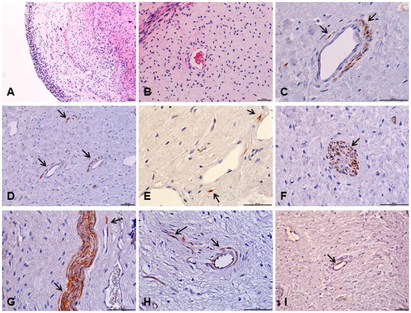
Figure 1.
Histological sections from dental pulp tissue stained with hematoxylin and eosin or submitted to immunohistochemical reaction. (a,b) Pulp tissue with no evidence of inflammatory reaction. (c,d,e,f) ALDH1 positive cells in human dental pulp. (g,h) CD90 positive cells. (i) STRO-1 positive cells. Stained cells were located mainly around the blood vessels and on the nerve structures of this tissue. Arrows indicate the immunoreactive cells. Scale bars: 50 μm.

