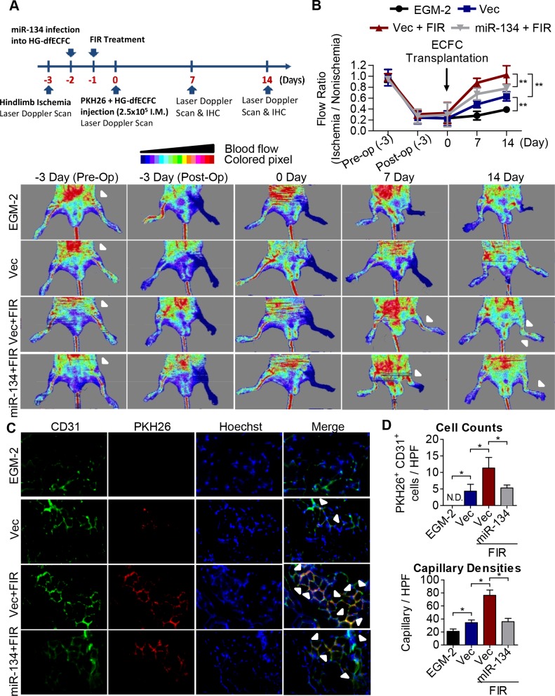
Fig 7. FIR induces ECFC activation and improves blood perfusion using an ischemic hindlimb model.
(A) Schematic representation of experimental design. (B) Upper: quantitative analysis of blood flow expressed as perfusion ratio of the ischemic to the non-operated contralateral hindlimb. n = 6, ** p < 0.01 by one-way ANOVA followed by Tukey’s post-hoc test. Lower: representative images of mouse ventral side during the measurement of hindlimb blood flow by laser Doppler before operation (Pre-Op), immediately after hindlimb ischemia surgery (Post-Op), and 2 weeks after intramuscular injection of culture medium (EGM2), HG-dfECFCs (Vec), HG-dfECFCs with FIR treatment (Vec + FIR), and miR-134 overexpressed HG-dfECFCs with FIR treatment (miR-134 + FIR). (C) Immunofluorescence staining of the tissue from nude mice 7 days after injection with PKH-26-labeled HG-dfECFCs. The capillaries in the limb muscles were visualized by anti-CD31 immunostaining (green), and injected human ECFCs were monitored by PKH-26 fluorescence (red). In addition there was Hoechst nuclear staining of the live cells (blue). (D) Quantitative analysis of CD31+/PKH-26+ double-positive cells and capillary densities in limb muscle of mice hindlimb ischemia region. HPF: high power field; N.D.: not detectable; * p < 0.05 by one-way ANOVA followed by Tukey’s post-hoc test.

