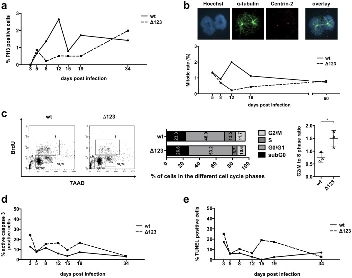
Fig 1. Phenotypic traits of B-cells infected with Δ123.
Primary B-cells were infected with B95-8 wild type (wt) or a recombinant virus lacking the three BHRF1 miRNAs (Δ123) and monitored between day 3 and 34 after infection. The mitotic rate was analyzed by immunofluorescence staining of phospho-histone H3 (PH3) (a), or Hoechst 33258 staining of DNA combined with a Centrin-2 (Cy3) and α-tubulin (Alexa488) double staining (b). BrdU incorporation assays were performed at day 13 to compare the cell cycle distribution of B-cells transformed with wt virus or the Δ123 mutant (c). The adjacent 2 graphs give the percentage of cells present in the different phases of the cell cycle as well as the ratio of cells in the G2/M and in the S phase, based on the results of the BrdU assay performed on three blood samples. We determined the degree of apoptosis in infected B-lymphocytes by immunofluorescence staining of active caspase 3 (d) or using a TUNEL assay at the indicated time points after infection (e). All experiments were repeated with cells from three different B-cell donors.

