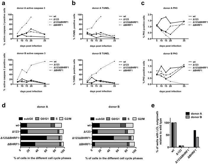
Fig 3. The reduced BHRF1 expression in B-cells infected with Δ123 leads to a moderately increased apoptosis.
Primary B-cells were infected with wild type, Δ123, ΔBHRF1 or Δ123ΔBHRF1 and monitored over time by immunofluorescence staining of active caspase 3 (a), by TUNEL assay (b), and by phospho-histone H3 staining (PH3) (c). Two out of five blood samples are shown. BrdU incorporation on day 14 was determined in B-cells transformed with wild type, Δ123, ΔBHRF1 and Δ123ΔBHRF1 (d). The results obtained with two samples are shown. Transformation assays were performed by infecting primary B-cells with 0.01 infectious virus per cell. 500 cells per well were seeded on 96-well cluster plates coated with 50Gy irradiated WI38 feeder cells and the number of wells with LCL outgrowth was determined 30 days after infection (e).

