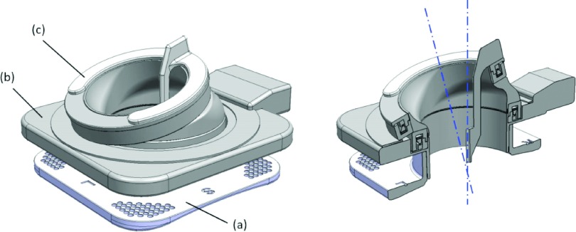
FIG. 2.
(Left) Model of the robotic instrument guide: (a) base stage with fiducial markers; (b) top stage with two ring actuation units; (c) detachable instrument guide. The instrument guide is detachable from the rotating units to accommodate multiple probe placements during image-guided cryoablation therapy. (Right) Cross-sectional model: the two axes of rotation created by the two ring-shaped actuation units. (Note that the intersecting point is placed on the bottom center of the device; in clinical applications, this placement will coincide with the skin entry point of the cryoablation probes.)

