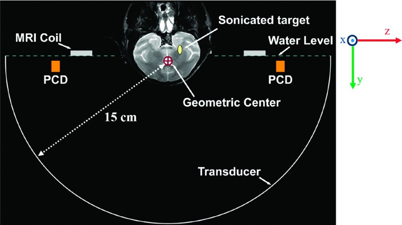
FIG. 1.
Coronal T2-weighted MR image of a macaque superimposed on a diagram of the experimental setup drawn approximately to scale. The locations of the MRI coil and PCD’s are indicated. The phased array transducer was used to electronically steer the focal point from the geometric center of the FUS array to a target near the skull base.

