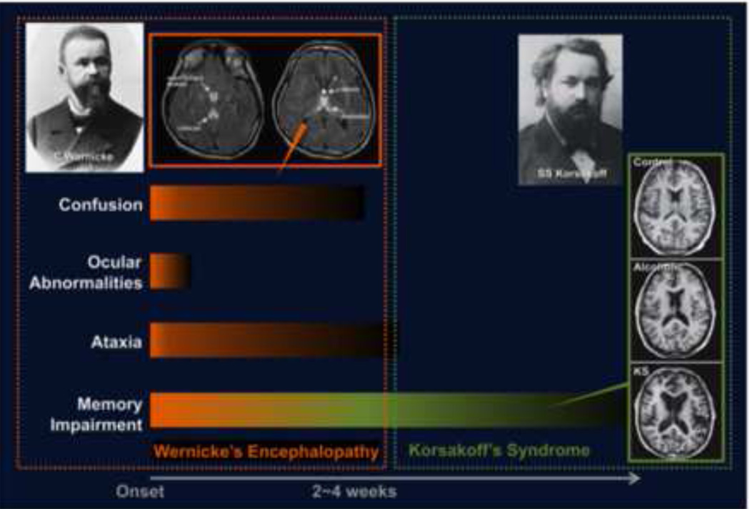
Fig. 1.
Depiction of the temporal progression of signs of Wernicke's Encephalopathy and its radiological signature of hyperintense areas, indicative of edematous tissue, in midline structures, and resolution to global amnesia and permanent structural damaged, most obviously marked by enlarged ventricular and sulcal spaces. Designed by Young-Chul Jung, M.D., Ph.D.

