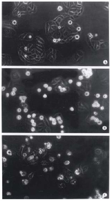
Figure 3.

Morphological changes of SMMC-7721 cells observed by light microscope after treatment with taxol for different times. (A) Untreated cells; (B) SMMC-7721 cells got round, diopter-enhanced after exposure to 10 nmol/L taxol for 24 h; (C) SMMC-7721 cells detached and blebbed after exposure to 10 nmol/L taxol for 48 h. × 400.
