
Figure 6.
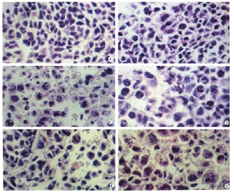
Histological changes of SMMC-7721 tumors after taxol treatment. (A) untreated control; (B) 2 mg/kg taxol, i.p. once daily for 10 d. Only slight morphological changes were presented; (C) 10 mg/kg taxol, i.p. once daily for 10 d. Most cells showed apoptotic changes and many apoptotic bodies were presented. Apoptosis were widespread. Apoptotic bodies were the small ovoid structures; (D) 24 h after i.p. injection of 10 mg/kg taxol. Mitotic arrest was marked; (E) 72 h after i.p. injection of 10 mg/kg taxol. Mitotic arrest could still be presented with more apoptotic cells; (F) 120 h after i.p. injection of 10 mg/kg taxol. Apoptosis was dominant phenomenon than ever before. Hematoxylin and eosin. × 400