
Figure 3.
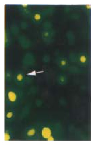
Cell slides under Nikon Eclipse TE200 fluorescence microscope using excitation light at either 488 nm to detect GFP (emission filter 522 nm). Distribution of GFP p27KIP1 fusion protein expressed in living HCC-9204 cells 8 after 100 μmol/L ZnSO4 treatment, indicated by an arrow. × 200