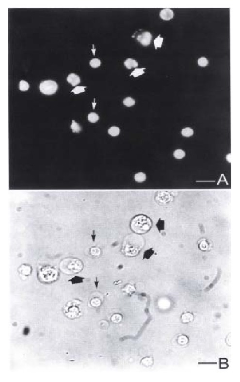
Figure 3.

Fluorescence and light micrographs of YAC-1 cells coincubated with pit cells at an E:T ratio of 10:1 for 3 h. (A) Fluorescence micrograph showing the apoptotic YAC-1 cells with fragmented nuclei (thick arrows) and pit cells ( thin arrows). Light micrograph shows the same field as (A). Bar = 5 μm.
