Figure 6.

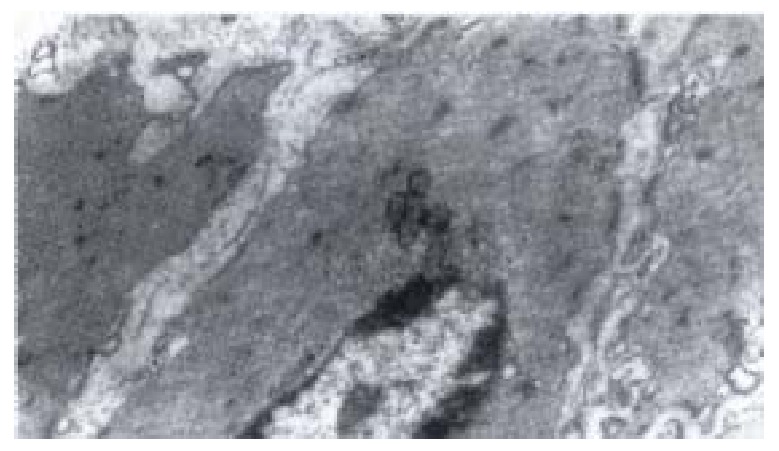
Electron-microscopic scanning of a control ‘s SO segment smooth muscle. The myofilaments were regularly arranged, kink mac ula densa clear and dense, plasmosome was normal.

Electron-microscopic scanning of a control ‘s SO segment smooth muscle. The myofilaments were regularly arranged, kink mac ula densa clear and dense, plasmosome was normal.