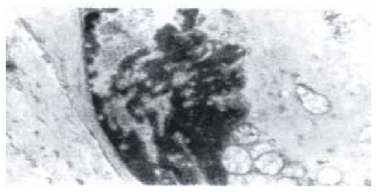
Figure 8.

Electron-microscopic scanning of a group II rabbit’s SO segment smooth muscle. It shows the swelling of plasmosome, irre gular arranged or fragmented intercristal space, some vacuolized, congregated at one end of nuclear, and disarragement of kink macula densa.
