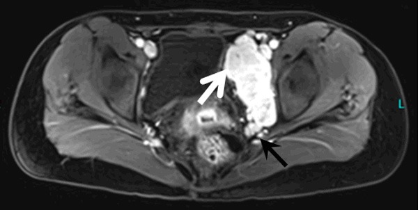
Figure 3.

MRI enhanced scanning indicates that the lesion is significantly enhanced in the left pelvic cavity, demonstrating a slight hyperintensity in the diffused-weighted images (white arrow), while a mildly enhanced hypointense area can be found in the upper part of the lesion. Thickened vascular imaging is observed below the lesion (black arrow).
