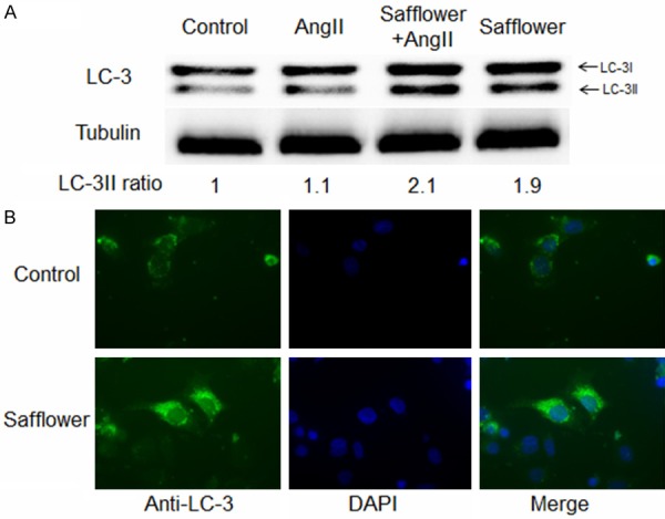
Figure 3.

Safflower extract induced autophagy in H9C2 cell. A. Western Blot analysis for LC-3II conversion. Differently treated cells was lysed and subjected to SDS-PAGE, then the separated proteins were transferred to PVDF membrane with anti-LC-3 antibody to detect the conversion of LC3-I to LC3-II. Quantification of image was conducted in QuantityOne program. B. Immunofluorescence assay for formation of autophagosome. Differently treated cells was fixed and penetrated by 2% paraformaldehydeand 1% Triton-X 100, respectively. The LC-3 antibody and FITC conjugated goat anti-rabbit IgG antibody were used to detection autophagosome formation.
