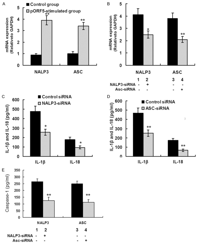
Figure 3.

pORF5 triggers IL-1β and IL-18 production via the NALP3 inflammasome. A. THP-1 cells were treated with pORF5 for 24 hours at 24 μg/ml. NALP3 and ASC mRNA expression was detected using qRT-PCR. The expression levels of the target genes were normalized to the endogenous control GAPDH expression and expressed as fold changes compared with the control group. The data represents mean ± SD of three independent experiments. Student’s t test. **P < 0.01. B. THP-1 cells were transfected with NALP3 (lane 2), ASC (lane 4), and negative control (lane 1 and 3) siRNA for 24 hours, then treated with 24 μg/ml pORF5 for an additional 24 hours. NALP3 and ASC mRNA expression was detected using qRT-PCR. The expression levels were obtained as the same in (A). The data represents mean ± SD of three independent experiments. ANOVA, *P < 0.05; **P < 0.01. C. THP-1 cells were transfected with NALP3 and negative control siRNA for 24 hours, followed by 24 μg/ml pORF5 treatment for 24 hours. Mature IL-1â and IL-18 protein expression was detected using ELISA. The data represents mean ± SD of three independent experiments. ANOVA, *P < 0.05. D. THP-1 cells were transfected with ASC and negative control siRNA for 24 hours, followed by 24 μg/ml pORF5 treatment for 24 hours. Mature IL-1â and IL-18 protein expression was detected using ELISA. The data represents mean ± SD of three independent experiments. ANOVA, **P < 0.01. E. THP-1 cells were transfected with NALP3 (lane 2), ASC (lane 4), and negative control (lane 1 and 3) siRNA for 24 hours, then treated with 24 μg/ml pORF5 for 24 hours. The conditioned supernatant was collected and caspase-1 protein expression was detected using ELISA. The data represents mean ± SD of three independent experiments. ANOVA, **P < 0.01 compared with control group.
