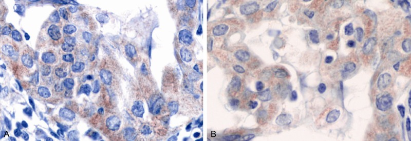
Figure 3.

Positive immunohistochemistry of MMP-2 and TIMP-2 in gastric cancer (A. MMP-2; B. TIMP-2). A and B shared the same judgment standard: the positive staining mainly aimed at the cytoplasm, with the peripheral interstitial partially colored; the evenly colored integrated cell was regarded as the positive cell.
